Achilles Tendon Diagram / 210 Achilles Illustrations Clip Art : Learn the basics of achilles tendon.. Diagram showing earthquakes and movement of the crust. Achilles tendon xenograft with medial and lateral aponeurotic fascial turndown flaps. The achilles tendon is a band of tissue that connects a muscle to a bone. Achilles tendon repair, operative technique. Learn about achilles tendon anatomy with free interactive flashcards.
Peroneal tendon tears and instability. Achilles tendon, strong tendon at the back of the heel that connects the calf muscles to the heel. You can see a diagram of the achilles tendon below. Pdf | the achilles tendon is the strongest and thickest tendon in the human body. With aging and overuse, the achilles tendon is subject to degeneration within the substance of the tendon.
A Multi Modality Approach Towards Elucidation Of The Mechanism For Human Achilles Tendon Bending During Passive Ankle Rotation Scientific Reports from media.springernature.com Your achilles tendons connect the muscles in your calves to the heel bones in your lower legs. It is also the commonest tendon to rupture. This most commonly happens during exercise or sport. With aging and overuse, the achilles tendon is subject to degeneration within the substance of the tendon. The tendon is formed from the gastrocnemius and soleus muscles. Achilles tendonitis causes & risks. This is in the calf, about two inches above the heel bone. It connects the heel to the large muscles of the calf and controls the movement of the foot.
With aging and overuse, the achilles tendon is subject to degeneration within the substance of the tendon.
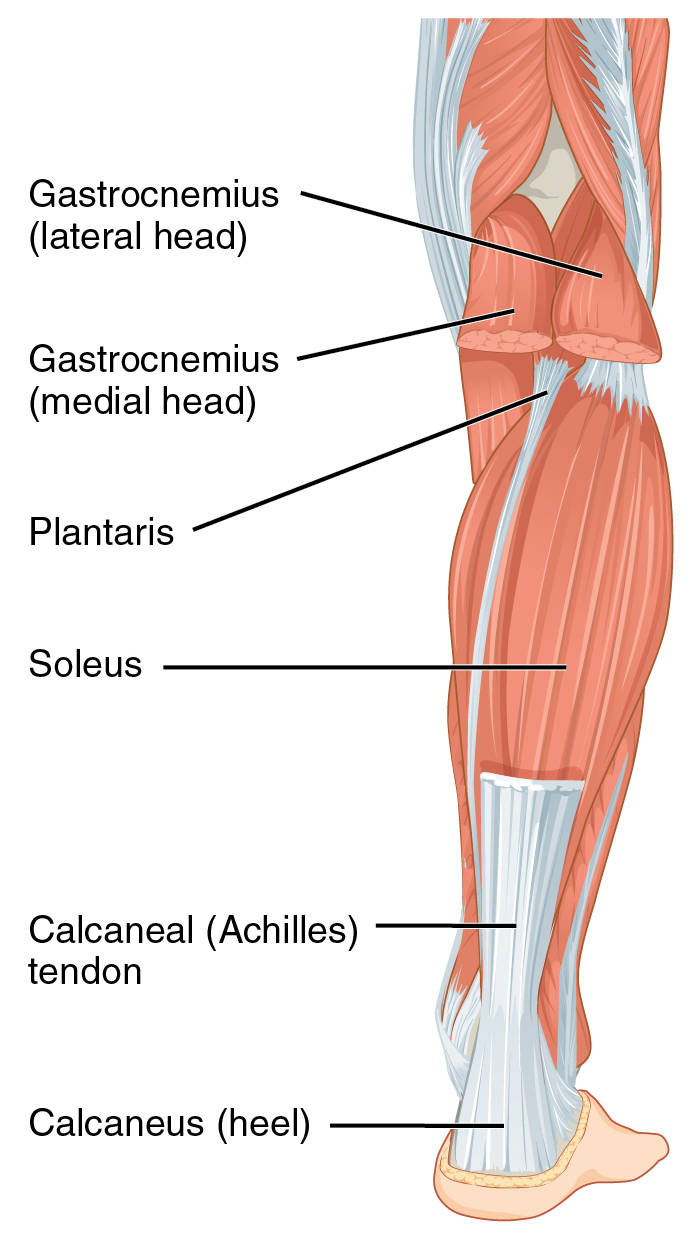
It serves to attach the plantaris, gastrocnemius (calf) and soleus muscles to the calcaneus (heel) bone. Achilles tendon, strong tendon at the back of the heel that connects the calf muscles to the heel. The achilles tendon is the thickest in the human body. By allison granot, mpt, ocs, cscs, palo alto medical foundation. It connects the two muscle groups (collectively, triceps surae) to the calcaneus. There is an area between the. Achilles tendon tears or ruptures the achilles tendon is the largest tendon in the body and the achilles tendon can tear or 'rupture'. The achilles tendon, seen in this diagram, attaches to the back of the heel bone (calcaneus) about halfway between the top and bottom of the back of the heel bone. Learn its anatomy and achilles tendon: Also called the heel cord, the achilles tendon facilitates walking by helping to raise the heel off the ground. The central movement it assists is the plantar flexion of your foot. Download this premium vector about diagram showing chronic achilles tendon tear, and discover more than 12 million professional graphic resources on freepik. It connects the heel to the large muscles of the calf and controls the movement of the foot.
The typical symptoms of this condition include localised achilles tendon pain that 'warms up' with activity. It connects the two muscle groups (collectively, triceps surae) to the calcaneus. The tendon is formed from the gastrocnemius and soleus muscles. This technique provides a description of achilles tendon reinsertion in the case of traumatic avulsion, as well as. Peroneal tendon tears and instability.
Achilles Tendon Wikipedia from upload.wikimedia.org It serves to attach the plantaris, gastrocnemius (calf) and soleus muscles to the calcaneus (heel) bone. Anatomy the achilles tendon is a strong tendon that connects the calf muscles to the heel. The achilles tendon is the large tendon connecting the two major calf achilles tendinitis is characterized by dull or sharp pain anywhere along the back of the tendon but usually close to the heel. The central movement it assists is the plantar flexion of your foot. The typical symptoms of this condition include localised achilles tendon pain that 'warms up' with activity. Also called the heel cord, the achilles tendon facilitates walking by helping to raise the heel off the ground. If the pain associated with your achilles tendon is severe or if you experience a sudden disability with. Download this premium vector about diagram showing chronic achilles tendon tear, and discover more than 12 million professional graphic resources on freepik.
Diagnosis an injury of the achilles tendon is a degenerative condition of the tendon, not an inflammatory process.
Learn the basics of achilles tendon. The central movement it assists is the plantar flexion of your foot. The achilles tendon or heel cord, also known as the calcaneal tendon, is a tendon at the back of the lower leg, and is the thickest in the human body. Download this premium vector about diagram showing chronic achilles tendon tear, and discover more than 12 million professional graphic resources on freepik. Insertional achilles tendonitis involves the lower portion of the heel where the tendon inserts to the heel bone. With aging and overuse, the achilles tendon is subject to degeneration within the substance of the tendon. The achilles tendon is the thickest in the human body. The achilles tendon, seen in this diagram, attaches to the back of the heel bone (calcaneus) about halfway between the top and bottom of the back of the heel bone. The achilles tendon is a band of tissue that connects a muscle to a bone. Your achilles tendon's function is to transfer the forces produced by your calf muscle to your foot. Rupture and discontinuity of the achilles tendon tendinous portions of the gastroc and soleus coalescing above… Diagnosis an injury of the achilles tendon is a degenerative condition of the tendon, not an inflammatory process. The tendon is formed from the gastrocnemius and soleus muscles.
If the pain associated with your achilles tendon is severe or if you experience a sudden disability with. Achilles tendonitis causes & risks. Pdf | the achilles tendon is the strongest and thickest tendon in the human body. This most commonly happens during exercise or sport. Learn about achilles tendon anatomy with free interactive flashcards.
Achilles Tendinopathy Exploring Injury Characteristics And Current Treatment Modalities Sciencedirect from ars.els-cdn.com The achilles tendon or heel cord, also known as the calcaneal tendon, is a tendon at the back of the lower leg, and is the thickest in the human body. The tendon is formed from the gastrocnemius and soleus muscles. The central movement it assists is the plantar flexion of your foot. Anatomy the achilles tendon is a strong tendon that connects the calf muscles to the heel. It is also the commonest tendon to rupture. The achilles tendon is the thickest in the human body. Hallux valgus, bunion in woman foot on white more similar stock illustrations. Learn its anatomy and achilles tendon:
Learn more from webmd about achilles tendon injuries, including their causes, symptoms, diagnosis, treatment, and prevention.
Peroneal tendon tears and instability. Anatomy the achilles tendon is a strong tendon that connects the calf muscles to the heel. It is therefore incorrect to describe this as tendinitis. The achilles tendon joins the calf muscles to the heel bone and runs down the back of the lower leg. This most commonly happens during exercise or sport. Achilles tendon repair, operative technique. Achilles tendonitis causes & risks. It is also the commonest tendon to rupture. The achilles tendon, seen in this diagram, attaches to the back of the heel bone (calcaneus) about halfway between the top and bottom of the back of the heel bone. Also called the heel cord, the achilles tendon facilitates walking by helping to raise the heel off the ground. The tendon is formed from the gastrocnemius and soleus muscles. Your achilles tendon's function is to transfer the forces produced by your calf muscle to your foot. Diagnosis an injury of the achilles tendon is a degenerative condition of the tendon, not an inflammatory process.
The achilles (calcaneal) tendon is a common tendon shared between the gastrocnemius and soleus muscles of the posterior leg tendon diagram. There is an area between the.


0 Comments